Tissues Science Chapter 6 | In-Text Questions & Answers | Exercises Questions & Answers | NCERT Class 9 Science | Notes | pdf
Q1. What is tissue?
Tissue is a cluster of cells which are structurally identical or similar and performs together a specialised function in the body. We learnt in previous chapter-5 that organisms are of two types – i) unicellular, & ii) multi-cellular. In case of unicellular organism (Amoeba, Bacteria etc.) all functions such as food intake, excretion etc. occur just inside a cell. But in case of multi-cellular organism (animals, plants) different cluster of cells perform different functions. In case of animals different tissues perform different specific functions in animals and plants. For example,
♦ Muscular tissue helps performing movement function.
♣ Blood tissue performs transportation of oxygen, hormones, food, waste materials etc.
♦ Nerve tissues performs as messenger.
♣ Vascular tissues in plants carry food from one corner to another corner.
Q2. What are the basic structural differences between animal and plant tissues?
Firstly, animals move from one place to another place for it’s livelihood. On the other hand plants are stationary. So animals require more energy than plants. Most of the animal tissues have living cell. Though plants are stationary but it grows up and requires supportive tissues that contain normally dead cells.
Secondly, plant growth is limited to certain regions where tissue division is happened lifetime. These tissue is called meristematic tissue. Other non-dividing tissues of plants are called permanent tissue. On the other hand animals have no specific dividing and non-dividing tissues as growth in animal is not limited to certain regions rather it has uniform growth.
Thirdly, various organ and organ system in animals are more specialised, specific and localised than plant. We find the basic differences between them in the ways of their life specifically in their feeding methods. Plant adapts itself for it’s sedentary existence whereas animals adapt themselves for it’s various activities.
Q3. Classify plant tissue.
We divide plant tissue into two groups i.e. a) meristematic tissue, & b) permanent tissue. Permanent tissue is further divided into two categories i.e. i) simple permanent tissue, & ii) complex permanent tissue.

Q4. Discuss meristematic tissue.
Plant growth is limited to certain regions where tissue division is happened. These tissues are known as meristematic tissue and theses are located in the growth regions of plants. We divide meistematic tissue into three i.e. i) Apical meristem, ii) Intercalary meristem and iii) Lateral meristem.
Apical meristem is located at tips of stem and roots where growing is happened and it results in increase in length of stem and roots.
Lateral meristem (cambium) causes increase in girth of stem and roots.
Intercalary meristem is located near the node in stem and roots. As it is growing part of plants, cell of this part has prominent nuclei, thin cellulose walls and dense cytoplasm but lacks vacuole. Characteristics of cells in growing parts remain same but slowly changes as they grow and mature with time and prominent difference is noticed with components of other tissues.


Q5. What is permanent tissue?
Cell in meristematic tissue gradually matures, stops division and gets permanent shape and size and turns into permanent tissue finally. The process from meristematic tissue to permanent tissue is known as differentiation. Further we divide permanent tissue into two groups. These are i) simple permanent tissue and ii) complex permanent tissue.

Q6. Discuss simple permanent tissue.
Simple permanent tissue exists below the epidermis. Details of various parts or section of simple permanent tissue are in the following –

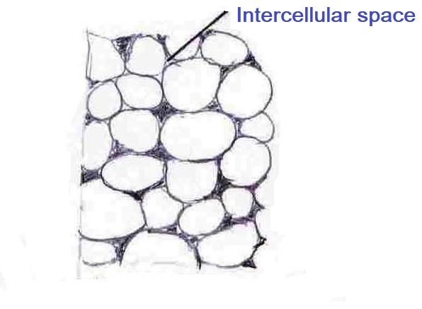
Parenchyma:
Cells in this parts stores food and are loosely arranged as inter-cellular space between them is more. Also these are living and have thin cell walls. Chlorenchyma is a kind of parenchyma which contain chlorophyll and takes part in photosynthesis process. Aerenchyma is also a kind of parenchyma which has large air cavity and helps aquatic plants to float.
Sclerenchyma:
This type of permanent tissue makes plant hard and stiff and gives strength to various parts of the plant. These type of tissue are present in hard covering of seeds and nuts, leaf veins, stems, in the region of vascular bundles etc. Cells in this parts are normally dead, long and narrow with thickened wall. Thickness of the cell wall is due to lignin and sometimes the cell wall is too thick to make internal space inside the cells in this permanent tissue.

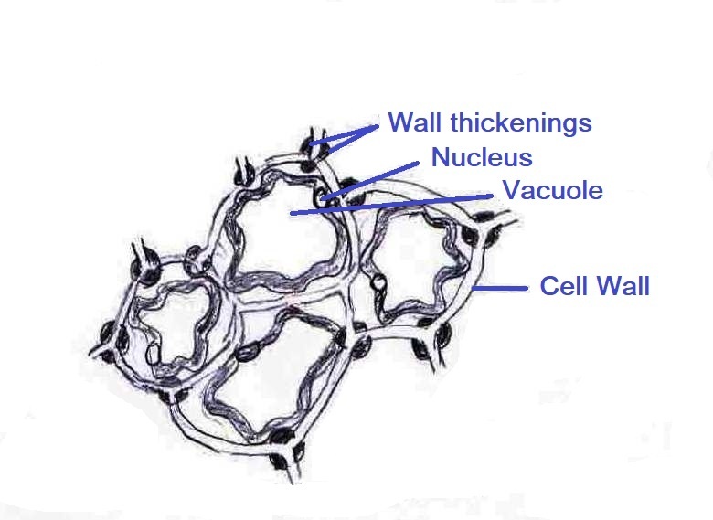
Collenchyma:
Cells in this parts add flexibility in plants and give mechanical support. Also they have very less inter-cellular space and help in bending of tendrils and stems to avoid breaking of plants. These cells are elongated and living with irregular thickened corners. You may notice this kind of permanent tissue in the leaf stalks just below the epidermis.
Epidermis:
Epidermis is the outermost single layer of cells and gives full outer covering to the plant. It protects plant from outside attack by the parasites and fungus. Epidermis also protects from mechanical injury. Cells in this permanent tissue are relatively flat with no inter-cellular space and form a continuous layer. Thickness of the outer layer and side walls of the epidermis are more than inner walls.

Desert plant has thick waxy and waterproof coating of cutin for avoiding of the loss of water as water loss in plants in the desert is very crucial for their survival and generally thickness of the outer layer is more in this case to avoid the loss of water. This tissue forms a water resistant, waxy and waterproof layer on the aerial parts of the plant to reduce loss of water. Stomata is the small pore in the epidermis of the leaf and is enclosed by two kidney-shaped cells. Transpiration and gaseous exchange happen through stomata. This tissue in roots has long hair-like parts to increase the surface area for facilitation of water absorption.

Q7. Discuss complex permanent tissue.
Single type of cell which are similar in nature forms single permanent tissue. But two or more type of cell which perform a common function constitute complex permanent tissue. Xylem and phloem are complex permanent tissue which mainly performs as a conducting or transporting tissue by making vascular bundle. These vascular tissues transport food and minerals which is essential for the survival of the plants in terrestrial environment.

Xylem:
It has xylem fibres, xylem parenchyma, tracheids and vessels. Xylem parenchyma stores food and xylem fibres perform supportive functions. Tracheids and vessels, which performs vertical transportation of water and minerals, are structurally tubular and have thick walls and many dead cells in mature condition.
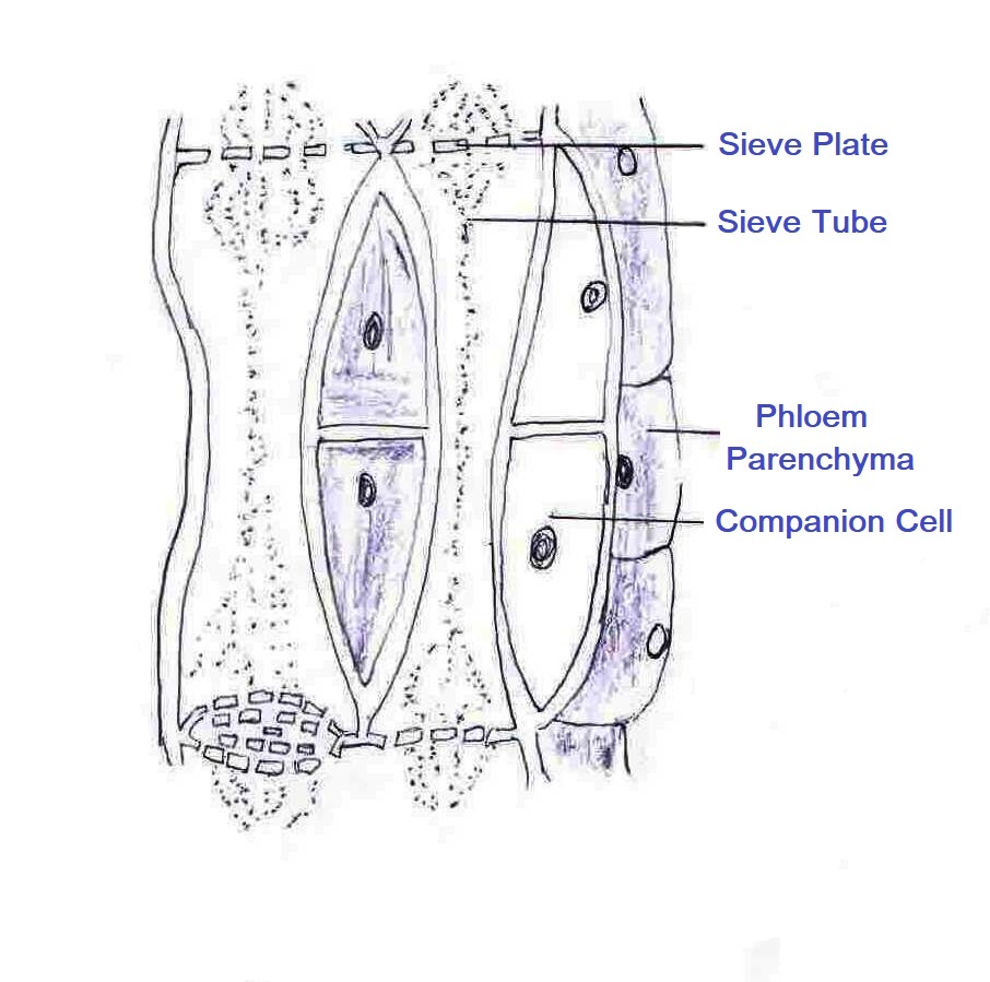
Phloem:
Phloem is consists of phloem parenchyma, phloem fibres, sieve tube, sieve cells and companion cells. Out of these five type cells, phloem fibres have dead cells and other type cells are living cells and sieve tubes are tubular cells having perforated walls. Main function of the phloem is to transport foods from leaves to other parts of the plant.
Q8. What are the various animal tissues?
Epithelial tissue, connective tissue, muscular tissue and nervous tissue are some important animal tissues.

Q9. Discuss epithelial tissue.
Epithelial tissues are the protective tissues in the animal body. It gives coverings to most organs and keeps them separate by forming a barrier. It also gives coverings to cavities within the body. Kidney tubules, the skin, lining of the mouth, lining of blood vessels, lung alveoli etc. are epithelial tissue. Cells in epithelial tissue are tightly packed and form a continuous sheet having no inter-cellular space but having small amount of cementing material between them.
As epithelial tissue gives covering, so any exchange between the body and outside environment or between various parts or organs within body must cross at least one layer of epithelium. So permeability of the cells plays a crucial role in regulating the exchange of material between different systems in the body. An extracellular fibrous basement membrane separates epithelium from the underlying tissue irrespective of any kind of epithelium.
Squamous Epithelium:
Different epithelial tissues located at various parts in the body have different type of structures in accordance with their unique function. Extremely thin and flat type cells form simple squamous epithelium.
Simple squamous epithelium forms a delicate lining. The epithelial tissues of the lining of blood vessels or lung alveoli in which transportation of substance happens through a selectively permeable surface are of squamous type.
The lining of the mouth, the skin of the body and the oesophagus are also examples of squamous epithelium. Epithelial tissues in the skin of the body exists in a pattern of layers to prevent wear and tear. Due to this type of pattern, it is also called stratified squamous epithelium.


Cuboidal Epithelium:
Cube-shaped cells form cuboidal epithelium. The lining of kidney tubules and ducts of salivary glands contain these type cells. Cuboidal epithelium mainly provides mechanical support. Sometimes it acts as gland cells and secretes substances at epithelial surface. Cells in this portion of epithelium form multicellular gland by folding inward. Due to this it is also called glandular epithelium.

Squamous Epithelium:
Tall and pillar-like (columnar) epithelial is located in the parts where absorption and secretion occur like in the inner lining of the intestine. It helps movement across the epithelial barrier. Ciliated columnar epithelium is located in the respiratory tract. Cells in this area have hair-like projections on the outer surfaces known as cilia. Cilia can move and it’s movement pushes the mucus forward to clear it.

Q10. Discuss connective tissue.
Loosely shaped cells which are embedded in an inter-cellular matrix, constitute connective tissue. Like the structure of the epithelial tissue (columnar, cuboidal, glandular etc), matrix of connective tissue may be rigid, fluid, dense or jelly type as per their their unique functions.

Blood:
Blood is an example of connective tissue. It flows and transports hormones, digested food, gases, waste materials etc. to different parts of the body. Matrix of blood is of fluid type where RBCs (Red Blood Corpuscles), WBCs (White Blood Corpuscles) and platelets are suspended. The fluid matrix of blood is known as plasma and it contains salts, hormones and proteins.

Bone:
Bone is also a strong and non-flexible connective tissue. It supports the main organs of the body, anchors the muscle and also forms the framework that supports the body. Matrix of bone is of hard type which is composed of phosphorus compounds and calcium compounds.
Ligament:
Ligament is also another connective tissue. It is very elastic and connects two bones with each other. Ligament has considerable strength and very little matrix. Tendons is a fibrous connective tissue with great strength and limited flexibility and connects muscles to bones.
Cartilage:
Cartilage is also a connective tissue. It smoothens bone surfaces at joints. Our nose, ear, trachea and larynx are soft due to having presence of cartilage tissue. Widely spaced cells in cartilage tissue forms solid matrix whose main constituents are sugars and proteins.

Areolar and Adipose:
It is also another connective tissue. Areolar helps in repair of tissues. It supports internal organs and also fills the space inside the organs. It is present in nerves, bone marrow, blood vessels and between the skin and muscles. Adipose is a fat-storing tissue and exits below the skin and between internal organs. Fat-storing helps adipose to act as an insulator. Cells forming adipose tissue are filled with fat globules.

Q11. Discuss muscular tissue.
Muscular tissue known as muscle fibres contains elongated cells. It causes movement in our body. It has special proteins known as contractile proteins. This proteins contracts and relaxes to cause movement.
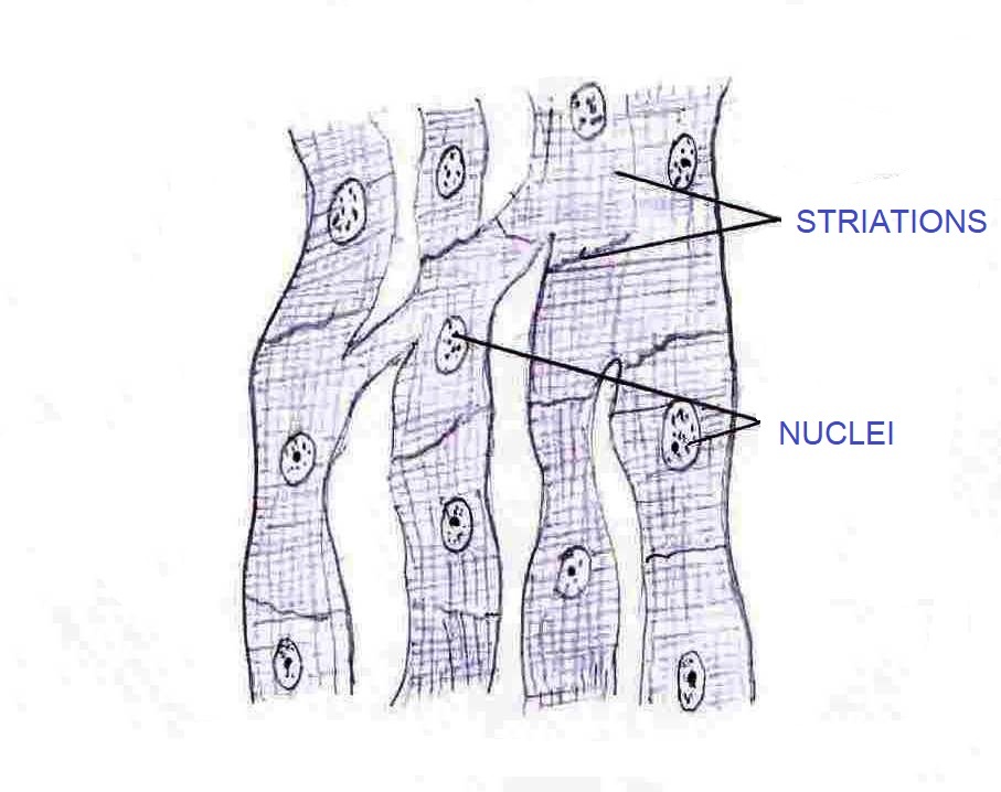
Voluntary muscles:
Voluntary muscles are the muscles that moves by our conscious will. Whenever we want, we can really start or stop their movement. For example muscles present in the limb of our body. These muscles are mostly attached to our bones and help in body movement. As it is attached to bones, so it is also called skeletal muscles. During microscopic analysis these muscles shows alternate light and dark bands or striations at stained conditions. For this reason they are also called striated muscles. Cells of this tissue are long, cylindrical, multi-nucleate and unbranched.


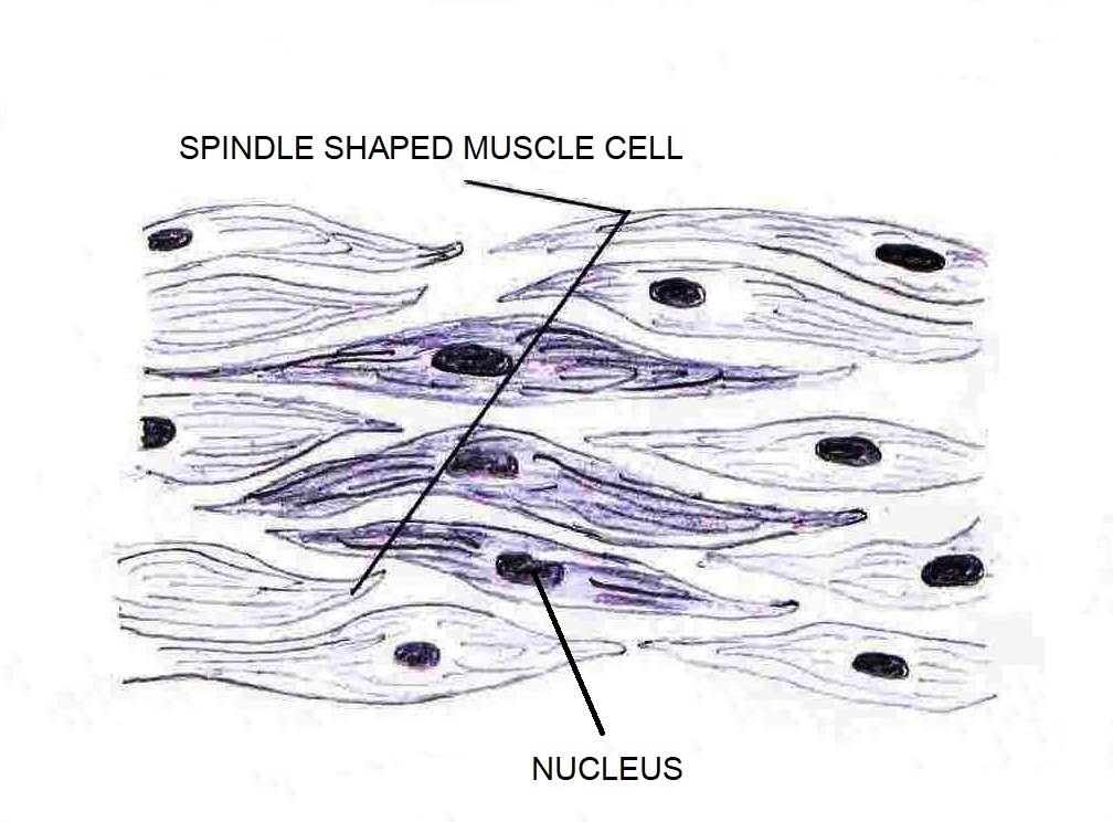
Involuntary muscles:
Involuntary muscles are those muscles whose movement do not depend on our conscious will. Whenever we want, we cannot start or stop their movement. For example – contraction and relaxation of blood vessels, movement of food in alimentary canal. Cells in this tissue are long with pointed ends and uni-nucleate. These muscles are also known as smooth & unstriated muscles. These are also present in the iris of the eye and in the bronchi of the lungs.
Another involuntary muscle is cardiac muscle that is present in our heart. Cardiac muscle continuously shows rhythmic contraction and relaxation throughout the life. Cells in this part are uni-nucleate, cylindrical and branched.
Q12. Discuss nervous tissue.
Nervous tissues are highly sensitive to any kind of stimuli. It transmits the stimulus very rapidly from one place to another pace in our body. It is present in our brain, nerves and spinal cord. Cells of this part are known as neurons. Cell body of neuron consists of a nucleus and cytoplasm from which hair like parts arise. Structurally a neuron may be up to a metre long and has a single long part (process) known as axon and many short, branched parts known as dendrites.
Lots of bounded nerve fibres constitute a nerve. A nerve impulse is a signal that passes through the nerve fibre and helps us to move our muscle by conscious will. Functions of nerve tissue and muscle tissue are interlinked, correlated and combined in such manner that it helps us to move rapidly in response to any stimuli.

In-Text Questions & Answers | NCERT Class 9 Science Chapter 6 Tissues
In-Text Questions & Answers | NCERT Class 9 Science Chapter 6 Tissues | Page No. 69
1) What is a tissue?
Tissue is a cluster of cells which are structurally identical or similar and performs together a specialised function in the body. For example – Epithelial tissue, connective tissue, muscular tissue & nervous tissue are animal tissues. On the other hand, meristematic tissue & permanent tissue are plant tissues.
2) What is the utility of tissues in multi-cellular organisms?
In case of unicellular organism, all the functions, such as movement, digestion & excretion are performed by single cell. On the other hand, a specific group of cell performs a specific function in multicellular organisms. This grouping allots various tasks to various groups of cells. For example, muscle cells form muscular tissues & epithelial cells form epithelial tissue. ‘Different tissue performs different specific function’ is the most important utility of tissues in multi-cellular organism.
In-Text Questions & Answers | NCERT Class 9 Science Chapter 6 Tissues | Page No. 73
1. Name types of simple tissues.
It is of three types i.e. – i) parechyma, ii) collenchyma, & iii) sclerenchyma. Cells of parenchyma are live, thin walled & relatively unspecialised. It forms the basic packing tissue. Cells of collenchyma are living, elongated & irregularly thickened. These cells have very little intercellular spaces. Collenchyma provides flexibility to plants. Cells of sclerenchyma are dead, long & narrow with thickened walls due to deposition of lignin. It provides rigidity & stiffness to the the plant.
2. Where is apical meristem found?
Apical meristem is located at tips of stem and roots where growing is happened. It results in increase in length of stem and roots.
3. Which tissue makes up the husk of coconut?
Cells of sclerenchyma are dead, long & narrow with thickened walls due to deposition of lignin. It provides rigidity & stiffness to the the plant. The husk of a coconut is basically mad up of sclerenchyma.
4. What are the constituents of phloem?
Phloem has four components. These are – i) sieve tubes, ii) companion cells, iii) phloem parenchyma, & iv) phloem fibres. Phloem mainly transports the soluble organic food prepared during the process of photosynthesis.
In-Text Questions & Answers | NCERT Class 9 Science Chapter 6 Tissues | Page No. 77
1) Name the tissue responsible for movement in our body.
Obviously it is muscular tissue. Skeletal muscles of muscular tissue are attached to bones. Their contraction make possible for locomotion, facial expression postures & other voluntary movements of the body. Read Q11 in detail.
2) What does a neuron look like?
Cells of nervous tissue are called neurons. Cell body of neuron consists of a nucleus and cytoplasm from which hair like parts arise. Structurally a neuron may be up to a metre long and has a single long part (process) known as axon and many short, branched parts known as dendrites.
3) Give three features of cardiac muscles.
a) Cardiac muscle is involuntary muscle. It is present in our heart.
b) Cardiac muscle continuously shows rhythmic contraction and relaxation throughout the life.
c) It does not get fatigue. Cells in this part are uni-nucleate, cylindrical and branched.
4) What are the functions of areolar tissue?
Areolar tissue supports many internal delicate organs. Areolar tissue also takes part in repairing of various tissues. It fills the spaces inside the organs. Therefore, it acts as a packing between the organs.
Exercises Questions & Answers | NCERT Class 9 Science Chapter 6 Tissues | Page Nos. 78 - 79
1. Define the term ’tissue’.
Tissue is a cluster of cells which are structurally identical or similar and performs together a specialised function in the body. In case of multi-cellular organism (animals, plants) different cluster of cells perform different functions. For example⇒
i) muscular tissue helps performing movement function.
ii) blood tissue performs transportation of oxygen, hormones, food, waste materials etc.
iii) nerve tissues performs as messenger.
iv) vascular tissues in plants carry food from one corner to another corner.
2. How many types of elements together make up the xylem tissue? Name them.
It has four elements i.e. xylem fibres, xylem parenchyma, tracheids and vessels. Xylem parenchyma generally stores food. Xylem fibres perform supportive functions. Tracheids & vessels performs vertical transportation of water and minerals. They are structurally tubular and have thick walls and many dead cells in mature condition.
3. How are simple tissues different from complex tissues in plants?
| Simple vs Complex Tissue | |
|---|---|
|
Simple Tissue |
Complex Tissue |
|
Same types of cells form simple tissues. |
Multiple types of cells forms complex tissues. |
|
Cells in simple tissues co-ordinate each other to perform a common function. |
Multiple types of cells in complex tissues co-ordinate each other to perform a common function. |
4. Differentiate between parenchyma, collenchyma & sclerenchyma on the basis of their cell wall.
| Parenchyma | Collenchyma | Sclerenchyma |
|---|---|---|
|
Cells of parenchyma has thin cell wall. |
In case of collenchyma, cell walls are thick at corners |
In case of sclerenchyma, cell wall is hard, rigid & very thick which reduces it’s cellular shape. |
|
Cell wall is made up of cellulose. |
Cell wall is made up of cellulose & pectin. |
It’s cell wall is made up of lignin. |
|
It has primary cell wall. |
Collenchyma has primary cell wall. |
Cell of sclerenchyma has secondary cell wall. |
5. What are the functions of the stomata?
The microscopic pores in the epidermis of the plant are called stomata. Two kidney shaped cells known as guard cells surround these pores. Stomata basically helps in the exchange of gases with the atmosphere. It also helps in transpiration which is the process of water loss in the form of water vapour.
6. Diagrammatically show the difference between the three types of muscle fibres.
There are three types of muscle in the body. These are –
a) Striated muscle. It is also known as skeletal muscle.
b) Unstriated or smooth muscle.
c) Cardiac muscle.
Draw pictures of a) Striated muscle, b) Unstriated, & c) Cardiac muscle given in Q11 to complete the answer.
| Striated Muscle | Unstriated muscle | Cardiac Muscle |
|---|---|---|
|
It is voluntary muscle. |
It is voluntary muscle. |
This is involuntary muscle. |
|
It has long cylindrical-shaped unbranched cells. |
This has spindle-shaped cells. |
It has short cylindrical-shaped branched cells. |
|
Cells are multinucleated. |
These cells are uninucleated. |
These cells are uninucleated. |
|
Fibres are arranged in form of bundles. |
Here fibres are arranged in form of sheets. |
Fibres are arranged in form of network. |
|
It has dark & light bands. |
Dark & light bands are absent. |
It has fainted dark & light bands. |
|
Locations – arms, legs, tongue, face neck, body. |
Locations – uterus, bronchi of lungs, alimentary canal wall, blood vessels, iris. |
Location – walls of heart. |
7. What is the specific function of the cardiac muscle?
Cardiac muscle is involuntary muscle. It is present in our heart. Cardiac muscle continuously shows rhythmic contraction and relaxation throughout the life. It does not get fatigue. Cells in this part are uni-nucleate, cylindrical and branched.
8. Differentiate between striated, unstriated & cardiac muscles on the basis of their structure & site/location in the body.
Write down answer of 6.
9. Draw a labelled diagram of a neuron.
Draw the picture of Q12.
10. Name the following ⇒
a) Tissue that forms the inner lining of our mouth.
Epithelial tissue.
b) Tissue that connects muscle to bone in humans.
Tendons.
c) Tissue that transports food in plants.
Phloem.
d) Tissue that stores fat in our body.
Adipose tissue.
e) Connective tissue with a fluid matrix.
Blood.
f) Tissue present in the brain.
Nervous tissue.
11. Identify the type of tissue in the following ⇒ skin, bark of tree, bone, lining of kidney tubule, vascular bundle.
Skin ⇒ stratified squamous epithelial tissue.
Bark of tree ⇒ cork tissue.
Bone ⇒ connective tissue.
Lining of kidney tubule ⇒ cuboidal epithelial tissue.
Vascular bundle ⇒ complex permanent tissue i.e. xylem & phloem.
12. Name the regions in which parenchyma tissue is present.
It is simple permanent tissue. It exists in leaves, fruits & flowers. Parenchyma tissue is basically involved in the function of storage & photosynthesis.
13. What is the role of epidermis in plants?
Epidemis is the outermost layer of cells in a plant body. Epidemis gives outermost covering & protects plant. It allows gaseous exchange from stomata. It also prevents water loss from the body of a plant.
14. How does the cork act as a protective tissue?
Dead cells form cork tissue. These cells are compactly arranged. They have no inter-cellular spaces. It has deposition of suberin which makes them impervious to gases & water. As a result cork tissue acts as a protective tissue.
15. Complete the following chart ⇒


Tissues Class 9 Science – To Be Continued – Tissues Class 9 Science